Some tissues absorb sound waves while others reflect them. What does prf mean in ultrasound.
 Wk 2 Liver Pathology Fatty Infiltration Diagnostic Medical Sonography Ultrasound Sonography
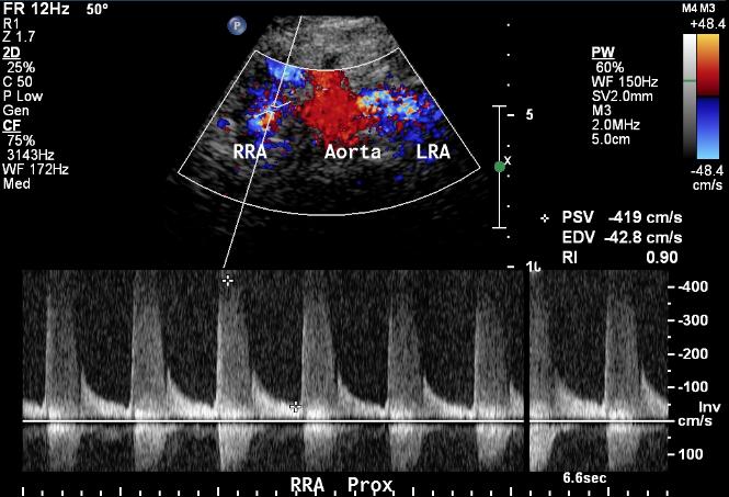
Color Doppler Duplex provides flow information within a region of interest ROI while spectral Doppler provides information within a sample volume.

What do the colors mean on an ultrasound. Do I Need A Test. Ultrasound screening has many advantages over contrast-enhanced CT and MRI such as accessibility low costs and no need for intravenous iodine contrast administration or. Colors on Abdominal Ultrasound.
Each of these represent the mean velocity within the region as measured by multiple PW sample volumes. My ultra sound of the liver showed dense liver. More intense color means the velocity is higher.
Each of these represent the mean velocity within the region as measured by multiple PW sample volumes. Various body tissues conduct sound differently. I had my right kidney removed in 211 for renal cell carcinoma.
Color doppler identifies moving velocities usually hues of red in one direction with respect to the transducer blue in the other usually used for identifying blood vessel flow. Color doppler of a simple cyst. Each of these represent the mean velocity within the region as measured by multiple PW sample volumes.
The use and contribution of color Doppler sonography for the assessment of pathologic entities in the neck is a method under clinical investigation. What do colors mean on a pelvic ultrasound. The color box is.
What the colors mean on a kidney ultrasound are the same as an ultrasound of the heart which we will discuss here. The color box is divided into small sample regions one color pixel. Red and Blue colors on an ultrasound refer to blood flow in the particular area.
Flow that travels away from the transducer negative Doppler shift is depicted in blue and flow that is traveling toward the transducer positive Doppler shift is depicted in red with lighter shades of each color denoting higher velocities. What do colors mean in thyroid ultrasound. 1 doctor answer 3 doctors weighed in.
Radiology 50 years experience. Blue means the blood is streaming away from the transducer and red means the blood is moving towards the transducer Note. What do i do when my ultra sounds comes out everything good.
RED means flow in one direction while BLUE means flow in the opposite direction. Do sounds in the knee mean I have arthritis. The test provides your doctor with important information about the flow of blood through your major.
What do the colors mean on a pelvic ultrasound. The different colors tell you in which direction the blood flow is going and can also tell the intensity or velocity of the flow. What do the yellow colors mean on an echocardiogram.
Most ultrasound images are in black and white but you can see differences in the shades of black and white in your ultrasound scan. The colors represent the speed and direction of blood flow within a certain area of the image color box. A Doppler ultrasound is a test that uses high-frequency sound waves to measure the amount of blood flow through your arteries and veins usually those that supply blood to your arms and legs.
The color on ultrasound is color Doppler. This device images frequency changes in the colors blue and red. What do white spots mean in an ultra sound.
Colour is blue or red depending on whether the blood movement is towards the ultrasound probe or away from it. Blue represents that the blood flow is away from the probe and red represents that the blood flow is towards the probe. One of the applications of Doppler ultrasound is color Doppler fig.
Blue and red does not necessarily mean low-oxygen and high-oxygen blood. The color differences come from the differences in the densities of the materials that the sound passes through. What do the colors mean on an ultrasound.
The two scans are identical the one on the right is outlined to help you understand what you are looking at. A third color usually green or yellow is often used to denote areas of high flow turbulence. It will not penetrate bone like an X-Ray.
The colors represent the speed and direction of blood flow within a certain area of the image color box. What do the colors mean in a color doppler ultrasound. The color Doppler shows normal vascularization of the.
The mean velocity is then converted into a specific color. The color box is divided into small sample regions one color pixel. How does a Doppler ultrasound work.
The lighter the shade of red or blue the more rapid and quick the blood flow. By definition flow towards the transducer is depicted in red while flow away from the transducer is shown in blue. Blood moving towards the transducer is red and blood moving away is blue.
Color Doppler sonography can Figure 1. When a renal mass is suspected conventional ultrasound and color Doppler imaging are often used for initial assessment. Depending on the angulations of the color box and the blood flow direction blood flow is displayed either in red or in blue.
CFM uses measurements of the velocity and direction of blood flow to superimpose a color pattern onto a section of a 2D image see Figure 4Traditionally flow towards the transducer is red flow away from the transducer is blue and higher velocities are shown in lighter shades. US-guided fine-needle aspiration biopsy of lymph nodes and tumors of the salivary glands is easy to perform and is characterized by high sensitivity and specificity. The above image is an ultrasound of a typical thyroid nodule except that this nodule is a bit bigger than usual.
The colors on the screen represent blood flow that is in motion. Ive had 3 CT scans with contrast since then but for todays test only an abdominal ultrasound was ordered. The colors represent the speed and direction of blood flow within a certain area of the image color box.
The colors on a kidney ultrasound represent the speed and flow direction of blood within a certain area known as velocity flow. The color box is divided into small sample regions one color pixel. Ultrasound is a non-invasive immediate tool used to image tissue.
So the first step to help you read the ultrasound image is to be familiar with the anatomy that you are imaging. A Grayscale image of a palpable breast mass that could be cystic or solid and B color doppler images demonstrate internal vessels thus this is a solid lesion. It detects movement and is mostly used to help evaluate blood flow in arteris and veins.
As the tech was scanning my left kidney the screen lit up with a lot of red and a small mount of yellow. The colors represent the speed and direction of blood flow within a certain area of the image color box.
 Pin On Breast
 Chorioangioma A Large Ovoid Mass Protruding From Fetal Surface Of The Placenta With Internal Calcifications Consistent With A Chori Sonography Placenta Fetal
 A Gallery Of High Resolution Ultrasound Color Doppler 3d Images Umbilical Cord Medical Ultrasound Ultrasound Ultrasound Tech
 Normal Anatomy Of Fetal Spine In Ultrasound Sonogram T 1059 Ultrasound Gynecology 4d Ultrasound
 Pin By Najlamohamed On Sonography Medical Ultrasound Ultrasound Technician Ultrasound Sonography
 Pin On Ultrasound Videos
 Ultrasound Basics How To Read An Ultrasound Image Ultrasound Diagnostic Medical Sonography Ultrasound Sonography
 Pin On Random
 Pin By Emily Van Haelen On Ultrasound Ultrasound Sonography Nurse
 Pin On Ultrasound
 Apical 2 Chamber Cardiac Sonography Ultrasound Sonography Student
 Color Flow Doppler Saturation Ultrasound Eca No Flow During Late Diastole Absent Color Flow Signal Vascular Ultrasound Medical Ultrasound Ultrasound
Color Doppler Flow Color Doppler Ultrasound Also Referred To As Color Flow Ultrasound Is Ultrasound Color Symbols
 Doppler Us Of The Liver Made Simple Radiographics Vascular Ultrasound Ultrasound Physics Radiology Imaging
 Pin On Vascular Ultrasound
 Pin On Cardiology
 6 Pitfalls To Accurate Lv Measurements Echo Echocardiography Cardioed Medical Ultrasound Cardiac Sonography Echocardiogram
 Normal Pancreas Ultrasound How To Ultrasound Diagnostic Medical Sonography Pancreas
 Pin By Denis Spaga On Radiology Medical Ultrasound Ultrasound Radiology